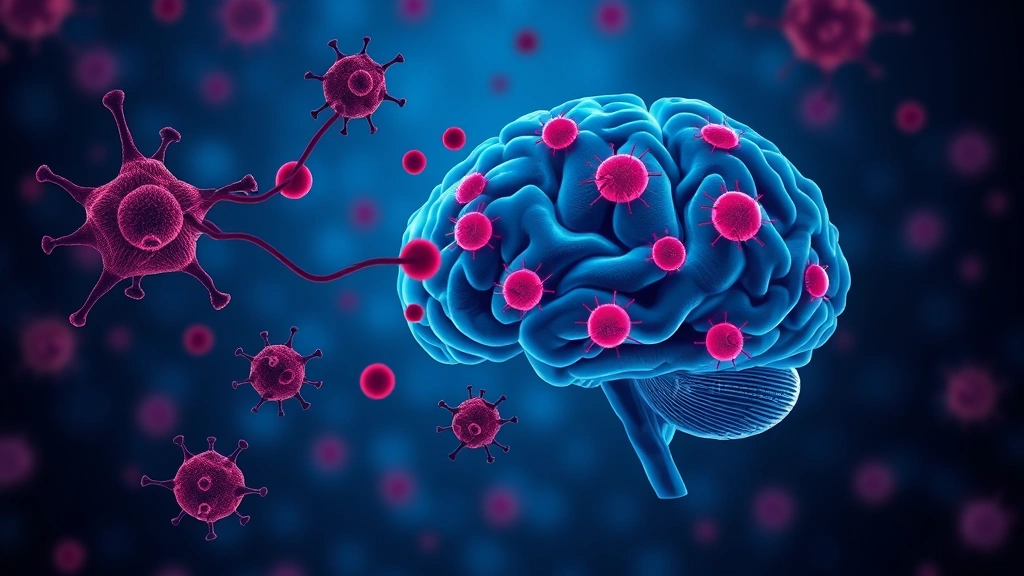
Immune cells that accumulate in the brain during Alzheimer’s-like disease and worsen the damage do not decide to attack on their own. They are activated in lymph nodes outside the brain — and interrupting that instruction protected memory in mice.
The work, from Washington University School of Medicine in St. Louis, was published in Nature Neuroscience as Priming of CD8+ T cells by peripheral dendritic cells exacerbates tau-mediated neurodegeneration.
Why immune cells are in this story at all
The brain was long considered immune-privileged — walled off behind the blood-brain barrier, largely invisible to the body’s immune surveillance. That view has eroded substantially. The central nervous system does drain to peripheral lymphatic tissue, meaning material from the brain can reach lymph nodes where immune responses are organised.
In parallel, evidence accumulated that neurodegeneration is not purely a matter of protein aggregates killing neurons. Immune activity appears to be an active participant, and in tauopathy — disease driven by the tau protein, the pathology that tracks most closely with cognitive decline in Alzheimer’s — T cells had been observed accumulating in affected brain regions.
What was unclear was whether those T cells were a cause, a consequence, or a bystander.
The relay the researchers traced
Studying mouse models of tauopathy, the team mapped a sequence with a clear division of labour.
Tau-damaged brain cells release material that reaches lymph nodes. There, classical dendritic cells type 1 (cDC1) flag that material as a target and prime CD8+ T cells — the cytotoxic subset that kills cells displaying whatever it has been trained to recognise. Those primed T cells then migrate into the brain, where they worsen the damage.
A striking detail: cDC1 cells within the brain itself do not appear to interact with these T cells. The activation step happens outside, exclusively. The brain receives armed cells; it does not arm them.
The experiment that makes the case
The team eliminated dendritic cells from the lymph nodes. Doing so wiped out the elevated T-cell levels in the brain and the associated damage — and the mice retained their cognitive abilities.
This is what separates the finding from an observation. Removing an upstream step and watching the downstream harm disappear establishes that the relay is necessary, not merely present. It also settles the causation question for these models: the T cells are contributing to damage, not simply arriving to inspect it.
Why the location is the point
The most consequential feature of this result is geographical.
The dominant obstacle in neurological drug development is the blood-brain barrier. Most molecules cannot cross it, which is why brain-targeted therapies resort to direct injection into cerebrospinal fluid, engineered transport mechanisms, or surgical delivery — all of which add cost, risk and complexity.
If the harmful process is initiated in peripheral lymph nodes, a drug targeting it never has to enter the brain. It only has to reach tissue that ordinary systemically administered drugs reach easily.
The available toolkit
“There are lots of ways to manipulate T cells that have been studied extensively” in other diseases, the researchers noted, but “many haven’t yet been explored for neurodegenerative diseases.”
That is the practical argument. Oncology and autoimmunity have produced an extensive pharmacology of T-cell modulation — drugs that block activation, prevent trafficking, or deplete specific subsets, many already approved with known safety profiles. If the relevant target in tauopathy is T-cell priming in the periphery, some of that toolkit may be repurposable rather than requiring invention from scratch.
What is not known
The researchers acknowledge that the specific trigger activating the dendritic cells remains unknown — what exactly the damaged brain releases, and what the T cells are being primed to recognise. That gap matters, because a therapy that broadly suppresses T-cell priming would carry the infection and cancer surveillance risks familiar from immunosuppression. A targeted intervention requires knowing the antigen.
This is also mouse work, and mouse tauopathy models reproduce features of human disease rather than the disease itself. Neurodegeneration research has a long record of interventions that protected mouse cognition and did nothing in people.
How this fits the shift away from amyloid
The finding lands in a field that has been broadening its view of what drives neurodegeneration.
For decades Alzheimer’s research concentrated overwhelmingly on amyloid, and the anti-amyloid antibodies that eventually reached the market produced real but modest slowing of decline — enough to validate the target, not enough to satisfy anyone. That gap pushed attention toward tau, which tracks more closely with cognitive symptoms, and toward the possibility that other processes contribute substantially.
Immune involvement is the most active of those alternative threads. Genetic studies have repeatedly implicated immune-related genes in Alzheimer’s risk, and microglia — the brain’s resident immune cells — are an established focus. What this work adds is a role for the adaptive immune system, organised outside the brain entirely, which is a different and more accessible target than anything inside it.
What the study offers is a mechanism with an unusually convenient location, and a rationale for testing an existing class of drugs against a disease they have never been aimed at. Preclinical research; not medical advice.